creation date: 2025-06-20 20:06
tags: Pathologies
Pulmonary Embolism
Background
Definitions
Pulmonary embolisms are defined as obstruction(s) of the pulmonary artery or one/multiple of its branches by thrombus, tumour, air, or fat.
Most commonly though, PE refers to those formed by thrombus and as such as a form of venous thromboembolism which is the form discussed here.
Etiology
In most cases, thrombus causing PE originate as lower extremity deep vein thrombosis (DVT) and thus causes reflect those of DVT. A number of factors predispose DVTs and PEs and are discussed in the risk factor section below.
In addition to risk factors, triggers for VTE include infection and malignancy (esp. pancreatic, hematological, lung, gastric, and brain). Other causes include myocardial infarction and congestive heart failure.
Pathogenesis
PE occurs when thrombus enters the pulmonary circulation. In the case of a lower extremity DVT, a blood clot breaks off and returns to the heart via venous circulation and enters the pulmonary artery. Fat emoblus can occur following large bone fractures.
Emboli more frequently affect the lower lobes than the upper. Larger emboli can obstruct the main pulmonary artery and cause saddle embolus.
Embolism results in impaired gas exchange due to reduction in perfusion and thus mismatch in ventilation-to-perfusion ratio. This leads to dead space ventilation and hypoxemia.
Unaffected branches may be affected due to vasospasm from mediators such as serotonin. and local accumulation of inflammatory mediators stimulates respiratory drive (tachypnea) results in hypocapnia and respiratory alkalosis.
Pulmonary hypertension may occur if occlusion is >30-50% of total pulmonary arterial bed. This results in increased right ventricular afterload and subsequent impeded outflow and dilation, and eventually hemodynamic instability.
Clinical Presentation
Signs & Symptoms
Presentation can vary significantly from asymptomatic to shock/sudden death.
Common features include:
- Dyspnea at rest or with exertion
- Pleuritic pain
- Cough
- Orthopnea
- Calf or thigh pain and/or swelling
- Wheezing
- Hemoptysis
- Hoarseness (dilated pulmonary artery)
Other signs found on examination include:
- Tachypnea
- Tachycardia
- Crackles
- Decreased breath sounds
- Accentuated pulmonic component of second heart sound
- JVP distension
- Transient or persistent arrhythmias
- Presyncope or syncope
- Acute shock
- Pleural effusion
- Fever
History & Physical Exam
Evaluation must be done vigilantly for PE as up to 1/3 of patients do not have mild or absent symptoms. Clinical suspicion should be investigated with assessment of risk factors.
Assess for shock and hemodynamic instability in cases of suspected PE.
Risk factors
The risk factors for PE are summarized by the Virchow triad: hypercoagulability, venous stasis, and endothelial injury.
Risks can be separated into genetic (Thrombophilia) or acquired which includes:
- Immobilization for prolonged period (eg. bed rest >3d, travel >4h)
- Recent orthopedic surgery
- Malignancy
- Indwelling venous catheter
- Obesity
- Pregnancy
- Cigarette smoking
- Oral contraceptive pill use
Other predisposing factors for VTE include:
- Lower limb fracture
- Major trauma
- Hip/knee replacement
- Congestive heart failure or respiratory failure
- Chemotherapy / cancer
- Central venous lines
- Postpartum period
- Infection
- Past history of VTEs
Diagnosis
Criteria
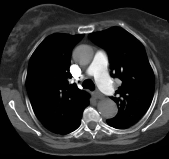
Diagnosis of PE is made by CT pulmonary angiography. Decision to undergo CTPA can be made using risk stratification criteria (see work-up). CTPA is both sensitive and specific for PE and provide information on location, prognosis, thrombus load, and right ventricular enlargement.
Indeterminant CTPAs may require a repeat (eg. if contrast bolus was poorly timed)
CTPA findings
PE will manifest as filling defects or an abrupt cutoff in a branch within the pulmonary vasculature. This can also manifest as the polo mint sign (ring of contrast around central filling defect) if artery viewed in the axial plane.
If CTPA is unavailable, a V/Q scan can be done. Findings are used with Wells criteria for diagnosis.
Work-up
In cases of hemodynamic instability with high suspicion of PE, once stability is re-established, anticoagulant treatment may be initiated prior to definitive diagnosis.
In stable patients, the work-up algorithm is as follows:
Determine pretest probability
Can be done using clinical gestalt or using a validated score such as Wells score, modified Wells score, or modified Geneva score. The Wells score stratifies risk into three categories seen in next step. Points are given as per MDCalc:
- Clinical signs/symptoms of DVT (+3)
- No better or equally likely diagnosis (+3)
- Heart rate >100 (+1.5)
- Immobilization at least 3 days OR surgery in past 4 weeks (+1.5)
- Previously diagnosed PE or DVT (+1.5)
- Hemoptysis (+1)
- Malignancy with treatment in last 6 months or palliative (+1)
PERC rule-out
Wells score <2 and not hospitalized, the PERC rule can be applied to see if a d-dimer is necessary. A PE can be ruled out if NONE of the following are true:
- Age ≥ 50
- HR ≥ 100 bpm
- O2 sat on room air <95%
- Unilateral leg swelling
- Hemoptysis
- Recent surgery or trauma
- Prior PE or DVT
- Hormone use
D-dimer
Wells scores from 2-6 or those <2 but scored at least one of the PERC criteria, a d-dimer can be done.
- D-dimers <500 ng/mL can exclude PE
- D-dimers ≥500 ng/mL indicates diagnostic imaging
Note that d-dimer levels may rise with age, resulting in a reduced specificity for patients >50 years of age. Consider using an adjust adjusted d-dimer.
CTPA
Wells score >6 or those with a positive d-dimer should be assessed with CTPA. In cases of a high clinical suspicion but negative d-dimer, CTPA may still be considered.
CTPA protocol involves timed image acquisition following a bolus of IV contrast. In cases where patient with a history of iodinated contrast allergy or impaired kidney function (eGFR <30 mL/min), a risk-benefit assessment should be made and antihistamine premedication may be considered.
Differential
There are a number of differential diagnoses based on the presenting complaint and are discussed in their respective work-ups.
Of note, a number of diagnoses mimic PE closely such as:
Red Flags / Complications
Delayed management may lead to hemodynamic instability, right ventricular failure, and sudden death.
Management
Acute treatment
Initial support and empiric anticoagulation
Initial considerations include:
- Maintaining O2 sat ≥90% using low or high-flow supplemental oxygen
- Hemodynamic support using IV fluids and vasopressors as needed
Empiric anticoagulation may be considered based on the risk of bleeding, clinical suspicion, the patient’s hemodynamic status, and the timing of diagnostic tests.
Low-risk PE (anticoagulation)
For patients without right ventricular dysfunction or hypotension and without bleeding risk, treatment consist of anticoagulation. DOACs are typically preferred.
Initial options for agents include:
- Low molecular weight heparin
- Fondaparinux
- Unfractionated heparin
- Oral factor Xa inhibitors
Common treatment options:
- Monotherapy of oral factor Xa inhibitor with rivaroxaban or apixaban
- Sequential therapy using LMWH (eg. enoxaparin) x 5 days then oral factor Xa inhibitor (eg. edoxaban) or direct thrombin inhibitor (dabigatran)
- Monotherapy of LMW heparin subq if PO not feasible or CI
- UFH IV in inpatient setting (preferred for kidney failure and hemodynamic instability)
- Avoid rivaroxaban in women with heavy menstrual bleeding (or other mucosal bleeding) - low evidence but hematologist recommended
In cases where anticoagulation is absolutely contraindicated, a vena cava filter may be used to prevent recurrent emboli from entering the pulmonary circulation.
Intermediate-risk PE (anticoagulation + monitoring)
In this patient group, anticoagulation similar to the low-risk group is used. Additionally, close monitoring of oxygenation, BP, and HR changes should be don to avoid deterioration.
Consider consulting for thrombolytic therapy or thrombectomy.
High-risk PE (reperfusion therapy)
In unstable patients or who becomes unstable again despite anticoagulation, systemic thrombolytic therapy is indicated. If that is not feasible, then consider catheter-based procedures or surgical embolectomy.
Systemic thrombolysis consist of administering tissue-type plasminogen activator (tPA). Unfractionated heparin (UFH) is typically used prior to thrombolysis and discontinued immediately before and during the infusion of tPA.
Patient should be monitored for stability but minor bleeding during infusion is common.
Long-term management
Goal is to complete treatment of acute episode and prevent recurrence of VTE. Management typically consistent of at least 3 months of anticoagulant (similar protective effect as 12-24 months of anticoagulant; use beyond that is a individual decision weighed against risk of bleeding).
References
Tools / Guidelines
MDCalc - Wells Score PE
MDCalc - PERC
MDCalc - Wells Score DVT
Thrombosis Canada - VTE treatment