creation date: 2026-06-18 03:11
tags: Workups
Acute Vision Loss
Background
Acute vision loss is almost always an emergency and timely evaluation is crucial for minimizing poor patient outcomes.
Acute transient vision loss: less than 24 hours; also referred to as amaurosis fugax
Acute persistent vision loss: at least 24 hours
Pathophysiology
Acute vision loss can occur due to disruption in the function of any of the structures along the visual pathway. This pathway is divided into three components–media, retina, and neural visual.
Media
To achieve clear vision, light has to travel from the front of the eye to the back through the cornea, aqueous humour, lens, and vitreous humour to the retina. Anything that may block the light’s path can cause vision loss.
Retina
The retina detects light entering the eye and converts it to neuronal signals. The retina can be torn/detached, become edematous, or become ischemic.
Neural visual pathway
Conditions that affect the pathway from the optic nerve to the visual cortex of the brain can affect vision.
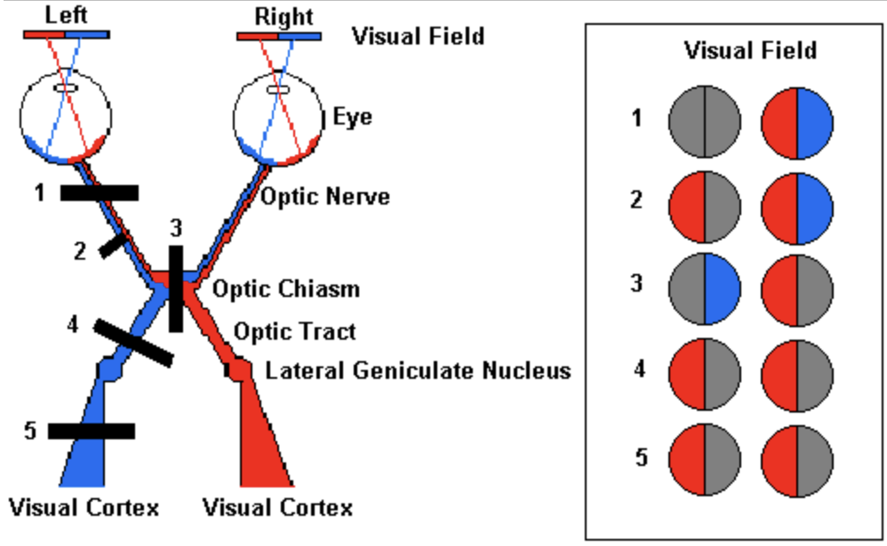
The visual pathway is summarized in this image:
Differential Diagnosis
Transient vision loss
Monocular:
- Ischemia
- Carotid artery disease
- Giant cell arteritis
- Cardiogenic embolism
- Hypotension
- Coagulopathy
- Retinal vein occlusion
- Retinal vasospasm and retinal migraine
- Optic neuropathy
- Papilledema
- Optic nerve compression
Binocular:
- Migraine aura
- Seizure
- Vertebrobasilar ischemia
Persistent vision loss
Conditions relating to the media:
- Corneal abrasion and keratitis (trauma, infection)
- Corneal edema (due to corneal abrasion, acute glaucoma)
- Hyphema (blood in anterior chamber; may be due to conditions causing growth or fragility of iris blood vessels such as diabetes)
- Lens changes (hyperglycemia, cataract/lens dislocation from trauma)
- Vitreous hemorrhage (trauma, spontaneous retinal tear, retinal neovascularization)
- Uveitis
- Ruptured globe (trauma)
Conditions relating to retina:
- Central retinal artery occlusion
- Central retinal vein occlusion
- Retinal detachment
- Acute maculopathy (eg. from trauma)
Conditions relating to neural visual pathway:
- Optic nerve lesions (monocular vision loss)
- Optic neuritis (most common in younger adults)
- Ischemic optic neuropathy (most common in older adults)
- Papilledema
- Orbital cellulitis (compression of optic nerve or direct infection)
- Chiasmal disorders (bitemporal hemianopia)
- Rapidly expanding compressive mass
- Infectious, vascular, inflammatory causes
- Trauma
- Retrochiasmal disorders (homonymous hemianopia)
- Stroke
- Hemorrhage
- Brain tumour
Initial Evaluation
History
Important elements that narrows differential:
- Recent ocular surgery (ophthalmologic referral)
- Contact lens use (consider abrasion, keratitis, corneal ulcer)
- Trauma (limited ddx)
- Pain
HPI should include:
- Exact acuity change; check monocular vision as chronic gradual loss of one eye may be mistaken for acute loss
- Laterality
- Quality
- Precipitating factors (eg. bright lights, posture, timing, eye movement)
- Redness
- Associated symptoms (neurologic deficits, systemic pains, GCA symptoms)
- Preexisting visual problems (including refractive status)
- Medical history of DM, CAD, HTN, hypercoagulability, or vascular risk factors
- Medication history
- Anticholinergics (accommodation loss, angle-closure glaucoma)
- Bisphosphonates (uveitis)
- Digoxin (yellow vision)
- Rifabutin (uveitis)
- Sildenafil (blue vision, ischemic optic neuropathy)
- Sulfonamides (myopia)
- Topiramate (angle-closure glaucoma)
- Oral contraceptives (ischemic, retinal, or optic nerve events)
- Fingolimod (macular edema)
- Cancer therapy drugs (retinopathy, uveitis, acute dry eye)
Physical Exam
Physical exam should include:
- Neurological exam
- Systematic eye exam (visual fields, pupillary response, EOM)
- IOP testing
- Slit lamp examination
- Fundoscopy
Investigations
Investigation may include:
- ESR/CRP (if GCA is suspected)
- Neuroimaging (if CVA is suspected)
In most cases, if an ophthalmologic cause is suspected, referral is warranted.