creation date: 2026-02-23 18:08
tags: Anatomy & Physiology
Spine
Vertebral Column
General Vertebral Anatomy
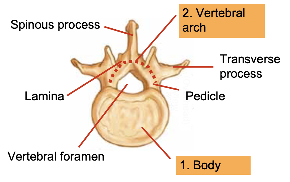
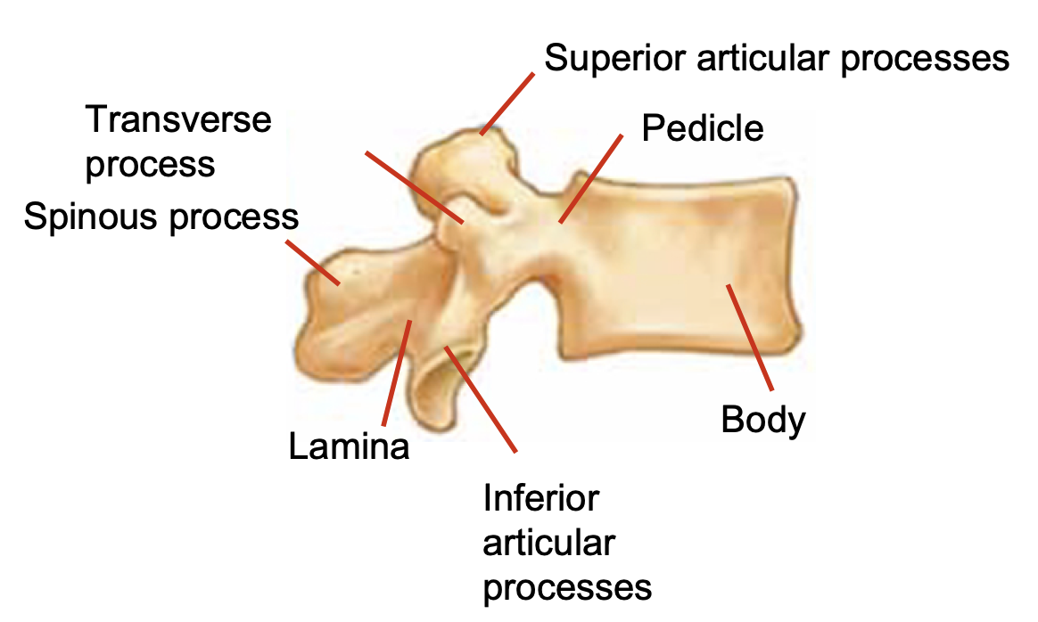
Each vertebrae stacks on top of the next, articulated with the inferior articular processes on the superior articular processes. This forms the zygapophyseal joint.
The vertebrae are stabilized by interspinous ligaments, anterior and posterior longitudinal ligaments, and supraspinous ligaments.
Between the body of each vertebrae are intervertebral disks. Each disk consist of lamellae on the outer edge, followed by anulus fibrosus moving inwards, surrounding a nucleus pulposus.
Cervical Spine
There are 7 cervical vertebrae (C1-C7). The c-spine has increased mobility and less weight bearing capabilities. The c-spine has approximately 20-40 degrees of AP curvature.
Upper cervical spine
The upper c-spine is specialized for supporting the cranium. It has large ROM for flexion, extension, and axial rotation.
C1: atlas
- No body
- 2 articular pillars and 2 arches allowing for large ROM and transition from c-spine to head
C2: axis
- Dens allows for rotation of C1 and C2 (embryologically C1 body)
- Poorly vascularized and osteoporotic
- Has foramen for vertebral artery
Lower cervical spine
The lower spine resists anterior translation and rotation. The vertebral artery transverses foramen in the transverse processes (except C7).
C7: vertebra prominens
- Refers to the prominent spinous process (useful for landmarking)
Thoracic Spine
There are 12 thoracic vertebrae (T1-T12). The thoracic spine has approximately 20-40 degrees of AP curvature in kyphotic alignment.
The thoracic spine is limited in movement and is relatively stable due to ribs and sternum. The spinal canal is limited in space, with the spinal cord taking up majority of the space.
Attached to the thoracic spines are the ribs:
- Rib 1-7 are true ribs (articulates with the sternum with costal cartilages)
- Rib 8-10 are false ribs (costal cartilages connect with rib 7 via costochondral joint)
- Rub 11-12 are floating ribs and do not articulate with the sternum.
Lumbar Spine
There are 5 lumbar vertebrae (L1-L5). The lumbar spine has approximately 40-60 degrees of AP curvature. The curvature transitions from kyphosis of the thoracic spine to lordosis.
Vertebral bodies are larger and lumbar spine is more mobile.
The conus medullaris lies at the level of L1 (but may be slightly higher at T12 or even as low as L3). This is the terminal end of the spinal cord in which the nerves become the cauda equina.
Sacrum and Coccyx
The sacrum consist of 5 fused vertebrae in a kyphotic curve. The coccyx (tailbone) consist of 3-5 vertebrae fused together.
Spinal Cord
The spinal cord is a cylindrical structure consisting of spinal nerves. It begins at the foramen magnum (as a continuation of the medulla oblongata) at the base of the skull and extends down the spinal canal of the vertebral column.
The spinal cord is divided into 31 segments (8 cervical, 12 thoracic, 5 lumbar, 5 sacral, and 1 coccygeal) which each has a pair of spinal nerves with their respective spinal root ganglia.
Within the spinal cord are both white and grey matter. The grey matter comprises of cell bodies while the white matter is axons (myelinated). There is less grey matter at the thoracic levels compared to cervical and lumbosacral.
The grey matter forms an H-/butterfly-shaped pattern which visually separates the various tracts.
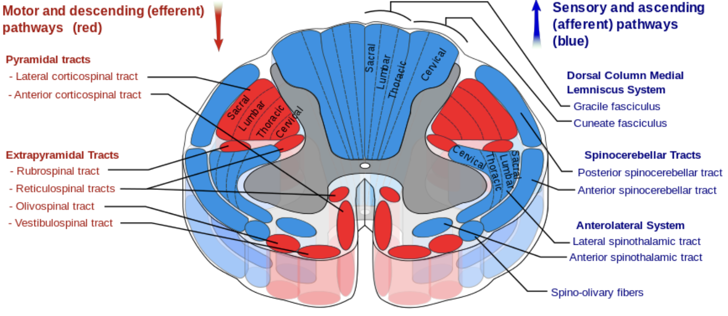
Ascending Fibre System
Dorsal Column / Medial Lemniscus System / Posterior Column
The dorsal column comprises of the laterally located fasciculus cuneatus (impulses from C1-T6) and medially located faciculus gracilis (impulses from T6 downwards).
The function of the dorsal column is:
- Fine sensation (two point discrimination)
- Vibration sense
- Prprioception
The axons for this tract originate as primary neurons from the ipsilateral dorsal root ganglion and travel upwards to synapse in the lower medulla.
The dorsal column is particularly susceptible to vitamin B12 deficiency.
Spinothalamic Tract / Anterolateral System
The spinothalamic tract consist of a ventral and a lateral component.
The ventral tract carries signals relating to:
- Crude touch
- Pressure sensations
The lateral tract carries signals relating to:
- Pain
- Temperature sensations
The peripheral axons reach the dorsal root ganglia and enter the spinal cord via the Lissauer’s tract which synapse within the dorsal horn and cross contralaterally after a short distance. The axons then ascend to synapse at the thalamus.
Spinocerebellar Tracts and Other Tracts
The spinocerebellar tracts are located at the lateral funiculus and carries unconscious proprioceptive information from muscle spindles and Golgi tendon organs to the cerebellum.
Other tracts include:
- Spinoreticular tract
- Spinoolivary tract
- Spinomesencephalic tract
- Spinohypothalamic tract
Descending Fibre System
Corticospinal Tract
This tract originates from the precentral motor cortex and includes the lateral corticospinal tract and the anterior corticospinal tract. These are known as the pyramidal tracts as they travel through the pyramids of the medulla. The nerves also cross at this time.
The lateral corticospinal tract is located medial of the posterior spinocerebellar tract and the anterior tract is located near the anterior median fissure.
The corticospinal tract innervates postural muscles and gradually shrinks towards the lower segments. The function of this tract is for limb and axial body movement and motor information for voluntary movement.
Extrapyramidal Tract
The extrapyramidal tract is primarily located in the anterior portion of the cord. Its role is to maintain changes in posture, muscle tone, and reflexes.