creation date: 2026-02-23 18:09
tags: Anatomy & PhysiologyIncomplete
Hand and Wrist
Skeleton
Connected to the radius and ulna are the carpal bones of the wrist. These form two rows and are represented by the mnemonic “So Long To Pinky, Here Comes The Thumb”
Thumb side — pinky side
| Trapezium | Trapezoid | Capitate | Hamate |
|---|---|---|---|
| Scaphoid | Lunate | Triquestrum | Pisiform |
Attached to the carpal bones are the five metacarpal bones. Each of which are attached the phalanges.
There are 14 total phalanges with three making up each finger (distal, middle, and proximal) and two making up the thumb.
Muscle and Ligament
Innervation
Sensory innervation:
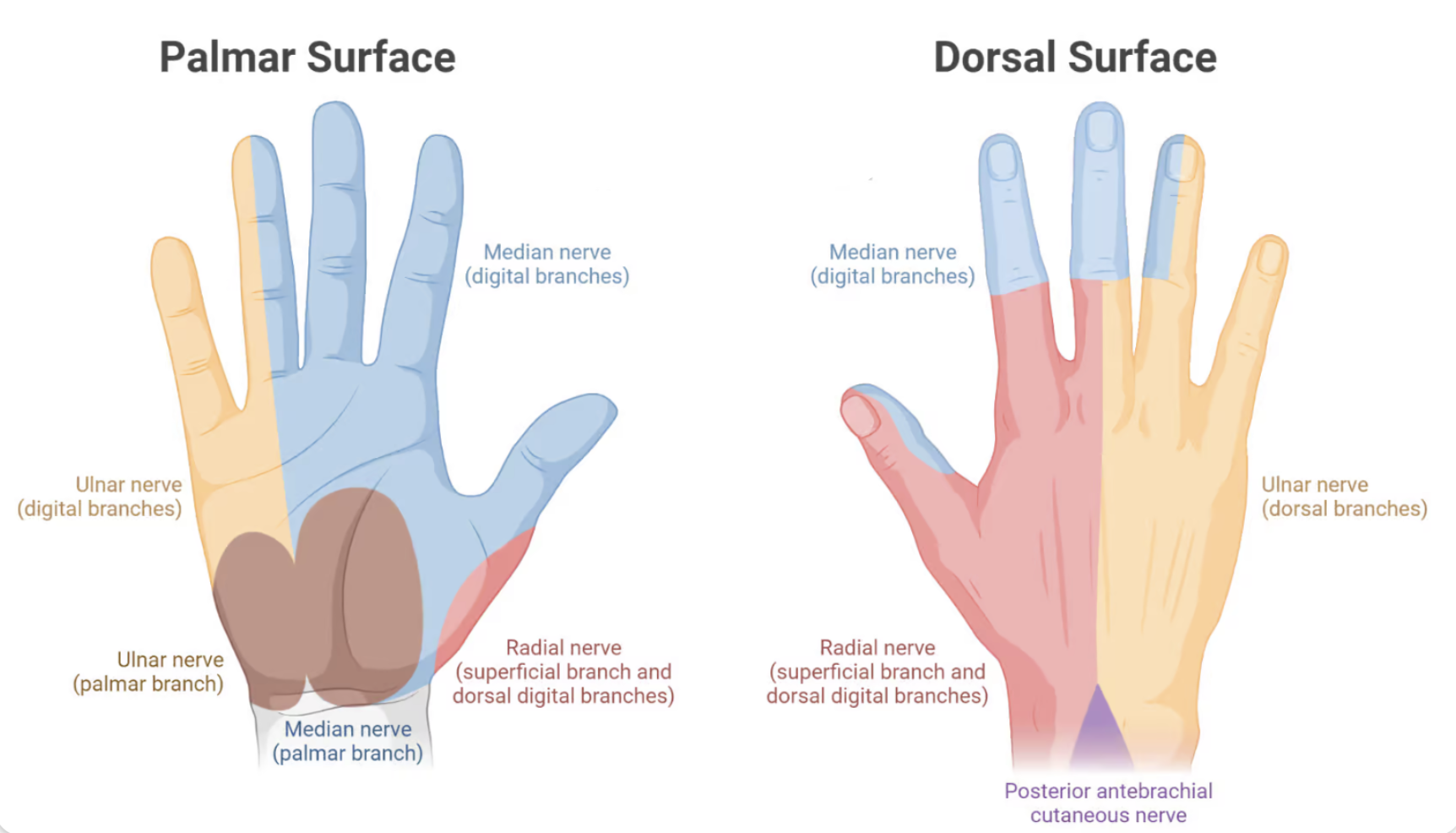
Radial Nerve
Motor function:
- Wrist extension
- Extensor carpi radialis longus/brevis
- Extensor carpi ulnaris
- Finger and thumb extension
- Posterior interosseous nerve (PIN) branch
Test function with wrist extension against resistance.
- Injury sign: wrist drop (inability to extend)
- Common injury site: spiral groove of humerus (“Saturday night palsy”) or radial head fracture
Median Nerve
The median nerve enters the hand through the carpal tunnel. Motor function are:
- Flexors (forearm)
- Flexor digitalis superficialis (PIP flexion for digit 2-5)
- Flexor digitalis profundus (DIP flexion for digit 2-3) via anterior interosseous nerve
- Flexor carpi radialis (flexion and radial deviation of wrist)
- Pronator teres (forearm pronation, weak elbow flexion)
- Pronator quadratus (forearm pronation)
- Thenar muscles (hand)
- Abductor pollicis brevis (thumb abduction)
- Opponens pollicis (thumb opposition; flexion, adduction, and medial rotation at CMC joint)
- Flexor pollicis longus (flexes thumb IP joint) via AIN
Test function with “OK” sign, make fist
- Injury sign: loss of thumb opposition and thenar wasting, ape hand (extension of digit 1-3 when trying to make a fist)
- Common injury site: carpal tunnel, supracondylar fracture
Ulnar Nerve
Motor function are:
- Flexion
- Flexor carpi ulnaris (wrist flexion, ulnar deviation)
- Flexor digitalis profundus (DIP flexion for digit 4-5)
- Lumbricals (MCP flexion, IP extension of digit 4-5)
- Flexor pollicis brevis (thumb MCP flexion)
- Abduction
- Hypothenar muscles (abduction, opposition, flexion of digit 5)
- Dorsal interossei (finger abduction)
- Adduction
- Palmar interossei (finger adduction)
- Adductor pollicis (thumb adduction)
Test function with observation at rest.
- Injury sign: claw hand (hyperextension of digit & flexion of digit 4-5 at rest), loss of abduction/adduction
- Common injury site: medial epicondyle (cubital tunnel) or hook of hamate